Article contributed by Julie Bokor (UF Center for Precollegiate Education and Training)
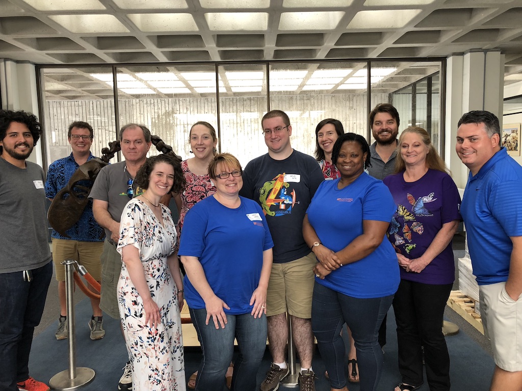
Using three-dimensional imaging as part of the openVertebrate project (or oVert) funded by the US National Science Foundation, scientists are creating digital specimens that can be viewed in the classroom, digitally dissected, 3D-printed, and more. In June, 2019 the oVert project partnered with nine secondary science educators to develop classroom lessons utilizing these digital specimens as part of the UF Center for Precollegiate Education and Training’s Summer Science Institute series: 3D Vertebrates, From Museum Shelves to Classrooms. Lead scientists and instructors were the oVert team of Dr. David Blackburn, Dr. Catherine Early, and Dr. Ed Stanley with additional students, faculty, and staff from the Florida Museum of Natural History.
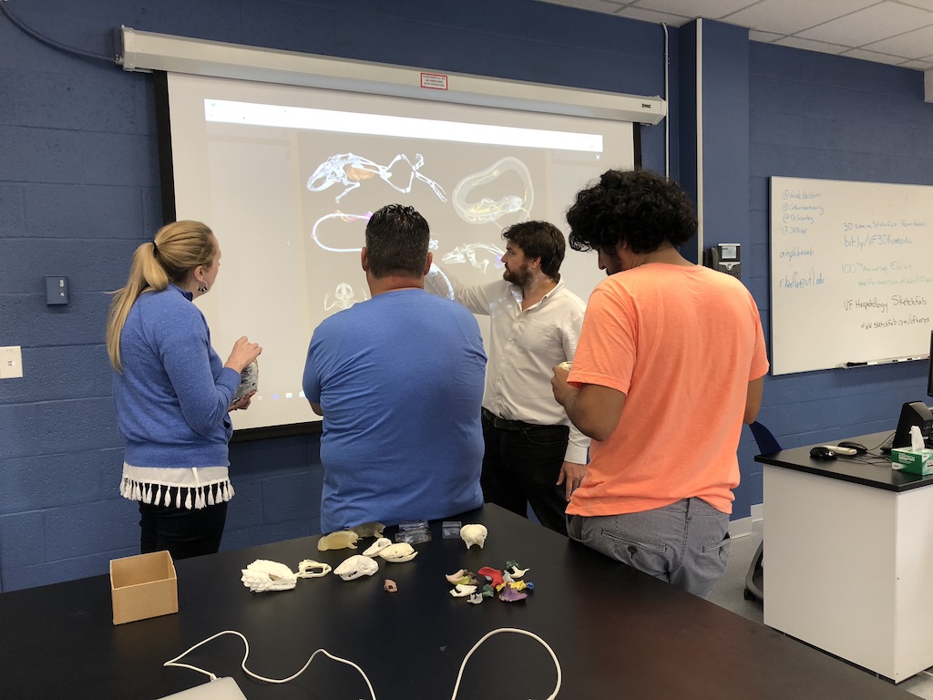 Following a general introduction to vertebrate diversity, participants learned how scientists and students at UF investigate anatomy, function, and evolution using digital three-dimensional anatomical data. During the week, brief presentations from and lunch time discussions with research faculty and graduate students highlighted the diversity of studies at the museum and UF as well as provided context to develop classroom lessons. Working in small groups with graduate and faculty mentors, participants selected a vertebrate species from the FLMNH’s herpetology and ichthyology collections and generate open-access 3D models based on data from CT-scanning and photogrammetry, adding to the oVert digitization effort.
Throughout the workshop, participants and scientists worked together to brainstorm ideas for translating the oVert project into classroom materials incorporating both 3D-printed and digital 3D objects and fully developed learning activities for implementation during the 2019/2020 school year. Learning activities are as varied as the educators and the students they teach. For example, one high school AP Biology lesson focuses on venom and explores concepts of evolution, protein structure and function, taxonomy and methods of scientific inquiry as students investigate a case study involving a young girl who has been bitten by an unidentified snake. Another lesson for 7th grade science integrates a study of joints using 3D printed bone segments to then develop a mechanical model that will allow movement in robots. The nine educators will continue to work with the oVert team during the school year as they implement their lessons and share their student learning outcomes and feedback for future use of digital specimens. More about the 3D Vertebrates, From Museum Shelves to Classrooms including the draft educator lessons can be found at: https://www.cpet.ufl.edu/teachers/ssi/ssi-2019/3d-vertebrates/.
The two Artec light scan generated models can be viewed on Sketchfab by clicking the links below:
Dermochelys coriacea (Leatherback sea turtle skull), UF-H-1422
Varanus komodoensis (Komodo dragon skull), UF 28219
The two CT-scanned specimens datasets are available in Morphosource and can be viewed by clicking below:


