 There was no dearth of enthusiasm or expertise at the recent Fluid-preserved Arthropod and Microscopic Slide Imaging workshop held at the University of Michigan September 16-18, 2013. For those whose interests span the gamut of cameras, lenses, microscopes, and the myriad gadgets and creative solutions that make it possible to capture images of difficult subjects, this workshop was the place to be.
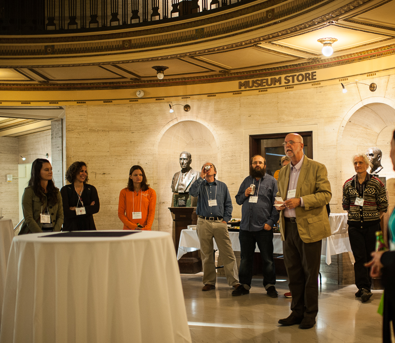
The impetus for the workshop originated with feedback from iDigBio's wet collections digitization workshop held at the University of Kansas in March 2013. This previous adventure focused on larger vertebrates, mostly fishes, with little attention given to small, difficult-to-image arthropods stored in vials...think spiders, millipedes, centipedes, and the like. The wet collections workshop also lacked effective strategies for imaging and recording data from microscopic slides, especially methods for capturing detailed representations of the organism under the cover slip.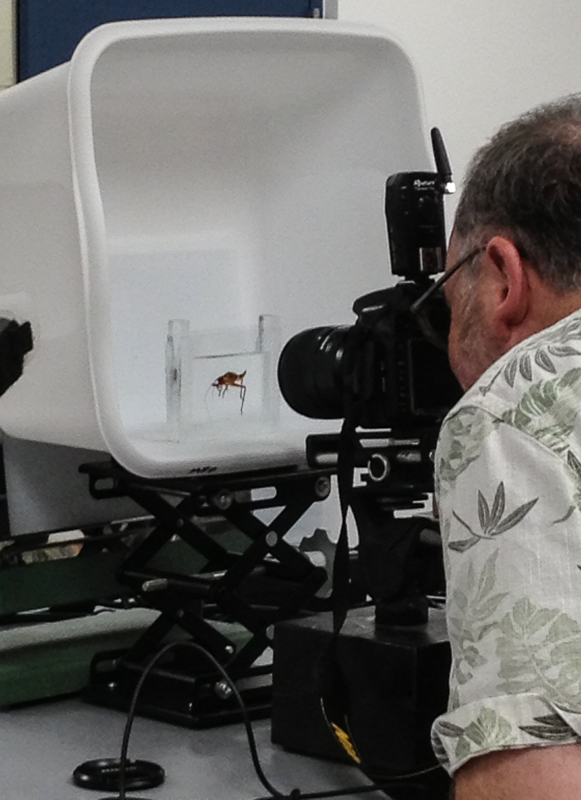
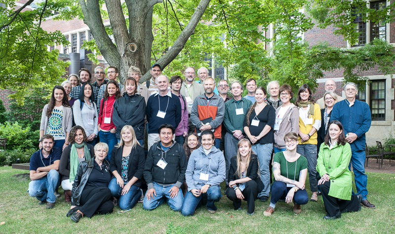
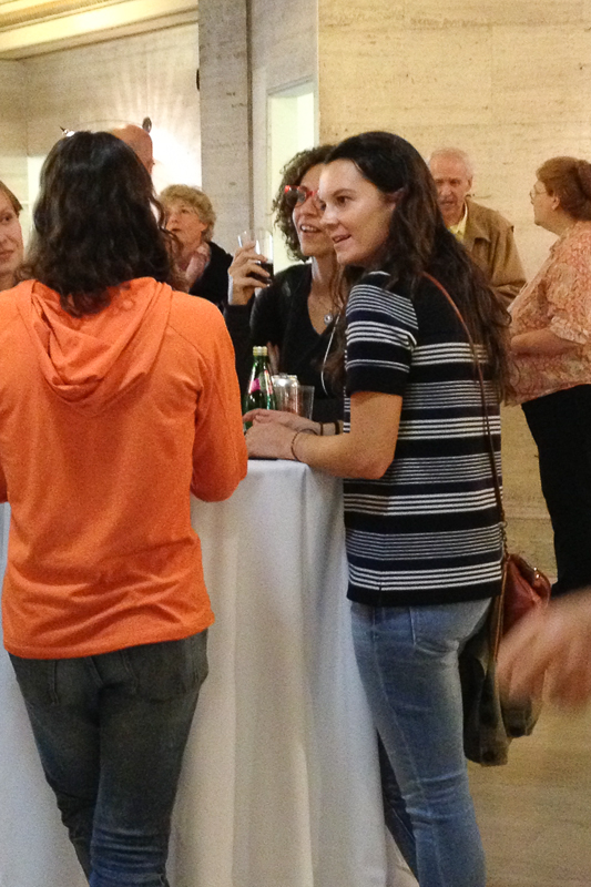
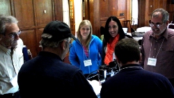
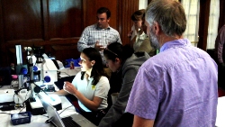
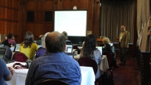
The content of the Ann Arbor wokshop included a host of excellent presentations and lots of outstanding discussion, all focused on imaging arthropods. Nearly 30 institutions were represented by the 40 participants, including long-distance travel winners Eirik Rindal and Karsten Sund from the Natural History Museum at the University of Oslo. Several afternoon demonstrations on both days of the workshop highlighted techniques, equipment, and novel imaging strategies, as well as the University of Michigan's newly renovated collections facilities. Participants also brought sample specimens to image during the demonstrations.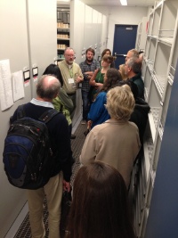
The workshop planning team included Mark O'Brien (U. Michigan), Sandy Brantley (U. New Mexico), Beka Baquiran (Field Museum), and Gil Nelson (iDigBio), and is commended for numerous contributions in time, expertise, and resources. Mark's onsite coordination coupled with his very informative demonstrations and presentations outlining successful, inexpensive strategies for arthropod imaging were particularly appreciated as were Sandy's observations and techniques for imaging arachnids. And, thanks to Beka for coordinating several excellent presentations from staff at the Field Museum.
 Special thanks also go to Dr. Diarmaid O'Foighill, director of the University of Michigan Museum of Zoology, for contributing the museum rotunda for the workshop reception and being on hand to welcome and inspire participants, and for his ongoing involvement in workshop activities.
The workshop was broadcast and recorded using Adobe Connect. The resulting videos are available on the workshop wiki for viewing, as are PDF files of the PowerPoint presentations.
It became clear to participants that a single, two-day workshop could not provide all of the answers to the perplexing problem of fluid-preserved arthropod imaging, which led to the establishment of a new iDigBio interest group designed to give more attention to this challenge. Workshop participants invite anyone interested in this topic to join this new initiative by contacting Gil Nelson (gnelson@bio.fsu.edu) and requesting to be added to the IDIGBIOINVI-L listserv.
 Given the complexity of the types of imaging explored, interest group members will continue the conversation in virtual meetings and future workshops. To this end, attendees prepared a summary document of issues related to fluid-preserved and microscopic slide imaging, all of which will become agenda items for future sessions. Important topics on this list include:
-
exploring the possibility of several, centralized slide-scanning centers to which institutions can send their material, the resulting image to be high enough quality to capture the organism as well as the slide label data,
-
establishing guidelines for prioritizing digitization targets,
-
creating guidelines for capturing standard views, including lists of important or key features by taxon,
-
finding ways to address image storage as a limiting factor when applying for or contemplating a digitization project, to include guidelines for computer configuration, network access, and cloud solutions,
-
motivating institutions to value and pursue digitization, utilizing iDigBio as a resource,
-
determining reasonable standards for image resolution,
-
defining suggested output sizes for different uses, e.g., thumbnails, feature amplification, systematics, and archive,
-
determining methods and strategies for increasing throughput,
-
establishing guidelines for determining background colors for given situations,
-
developing a matrix outlining imaging station specifications by purpose of project, expected outcome, and budgetary constraints, also to include notes about particular problems, successes, or features of each station,
-
studying the effects of hand sanitizer as a photographic medium to ensure its safe use,
-
determining whether imaging specimens held in vials is useful taxonomically without removing the specimen from its container,
-
exploring techniques for panoramic views of vial-stored specimens,
-
exploring strategies for imaging labels while still in the vial,
-
studying the use of vial imaging for inventory control and geo-referencng,
-
sharing and critiquing images contributed by interest group members,
-
exploring the possibility of partnering with Visionary Digital on a workshop at their lab.
The group is especially interested in item 17, above, and plans are in process to launch an image-sharing forum through which contributors can critique each other's images and suggest ways in which changes in lighting, technique, equipment, or other relevant parameters might be improved. An active listserv (IDIGBIOINVI-l@lists.ufl.edu) has been created to facilitate interaction and set the stage for future progress. Watch for more details on the workshop wiki.